Exercise physiology
VO₂max: what a single number doesn’t tell you
VO₂max sums up what the oxygen chain (lungs, heart, blood, vessels, capillaries, muscles and mitochondria) produces when it runs at full capacity. Understanding the workings of this integrated system makes it easier to identify what training should stimulate to make VO₂max “climb”.
Most of us roughly know the definition: VO₂max is the maximal rate at which the body can use oxygen during maximal exercise. In other words, it is the maximal amount of oxygen we are able to use per minute. It is a “ceiling”, a limit on oxygen uptake that the body cannot exceed, however intense the exercise. This limit varies greatly from one person to another: it is individual and depends on many factors. The good news: your VO₂max is dynamic, in the sense that it can change over time. It can be partly shaped by appropriate training that stimulates the various components underlying VO₂max.
But what really lies behind VO₂max? VO₂max is in fact a marker that integrates a complex reality… and the journey begins in the air around us. The oxygen it contains enters the lungs and crosses the wall of the alveoli into the blood, where haemoglobin binds it. The heart pumps this blood, the vessels distribute it to the active muscles, and it is in the capillaries, the finest of them, that oxygen leaves the blood to enter the muscle fibre. There, the mitochondria, small specialised structures inside cells, use it to regenerate ATP (adenosine triphosphate), the well-known molecule whose breakdown supplies the energy our muscle cells need to contract.
The number
VO₂max: the maximal output of an integrated system
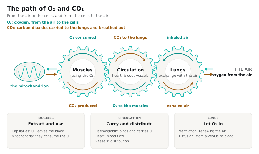
VO₂max is usually expressed in two ways. The absolute value is expressed in litres of oxygen per minute, whereas the relative value is expressed in millilitres per kilogram per minute. The latter relates the rate to body mass: it can therefore change with body weight, without any change in the absolute rate. It is often this relative figure that athletes like to compare.
The gold standard for determining VO₂max is an exercise test with analysis of respiratory gases. A mask channels inspired and expired air; the device measures the volume of air passing through and the concentrations of oxygen and carbon dioxide in that air. The difference between what goes in and what comes out gives the oxygen actually consumed and the carbon dioxide actually produced. What we observe is therefore the overall output of the chain (lungs, heart, blood, vessels, capillaries, muscles and mitochondria together), not the work of any one of these links taken separately [2].
During an incremental exercise test, oxygen uptake rises with intensity, supplying the mitochondria with more and more O₂ so that they can meet part of the rising energy demand. When the effort is maximal (and in fact slightly before), a saturation phenomenon may be observed: a plateau in oxygen uptake. This is the limit of what the body can consume per minute: VO₂max.
That said, a truly maximal effort is not always guaranteed during an exercise test: we therefore need to be able to judge whether the effort really was maximal. Strictly speaking, we should in principle speak of VO₂peak in an exercise test, because there is never any certainty that the body has actually reached its absolute maximum oxygen uptake: VO₂max. In practice, if several criteria are met (which must be analysed in detail), we can reasonably assume that VO₂peak does reflect VO₂max. Out of caution and intellectual honesty, we should always report VO₂peak or, at the very least, qualify what we call VO₂max [2,19].
Several things feed the uncertainty about whether VO₂max has really been reached.
- The effort itself. A test does not always continue to exhaustion. It may stop because the person decides so (flagging motivation, limited tolerance of discomfort or localised pain). The effort can therefore be interrupted before that ceiling is reached.
- The modality. The muscle mass engaged during exercise differs from one modality to another. As a result, so does oxygen uptake. The same person can therefore show different maximal values depending on the modality used for the test: often lower on a bike than when running [12]. The choice of modality is decisive when testing an athlete: otherwise, you obtain reference values that lose much of their meaning once transferred to another modality.
- The protocol. How the load increases also matters. On a cycle ergometer, the protocol can also influence the measured value, for example a ramp, where resistance increases continuously, or a step test. In a study by Michalik et al. [18], 57 young road cyclists (males, mean age 17 years) performed both tests one week apart, in random order: the ramp gave on average 65.8 mL·kg⁻¹·min⁻¹, versus 64.3 for the steps. The difference is small, but it is enough to show that the measured value also depends on how the test was conducted.
- The criteria are not neutral. To judge whether a test went far enough, we rely on a set of indicators: the ratio of carbon dioxide released to oxygen consumed (the well-known RER, Respiratory Exchange Ratio), heart rate, blood lactate and perceived exertion (the well-known RPE, Rating of Perceived Exertion). None is sufficient on its own, and the thresholds used must take into account factors such as age and sex [2,14].
- A plateau is not always observed. In a study by Day et al. [19], 71 men aged 19 to 61 performed a ramp test on a cycle ergometer, up to the limit of what they could tolerate. A plateau was observed in only 12 of them; in all the others, oxygen uptake kept rising until the test stopped. Yet the absence of a plateau does not prove that the maximum was not reached: in the same study, 38 participants performed, on another day, a constant-load effort at about 90% on average of the peak power reached in the ramp, up to the limit of tolerance, and their peak value was the same as in the ramp. Six of them also performed several constant-load efforts, all at very high intensity (above the level at which oxygen uptake can still stabilise), but at different loads. Whatever the load, their oxygen uptake levelled off at the same value (a plateau, clearly visible this time), and that value was the one reached at the end of the ramp [19]. This is the principle of the verification test, used mainly in research: a second constant-load effort, taken to the limit of tolerance, most often on the same day after 1 to 90 minutes of recovery and above the peak power of the test. In a meta-analysis of 54 studies, Costa et al. [15] found an average difference of only 0.03 L/min from the test. In other words, when the other indicators all point the same way, a true plateau is not essential [2,19].
A useful equation
The Fick equation, term by term
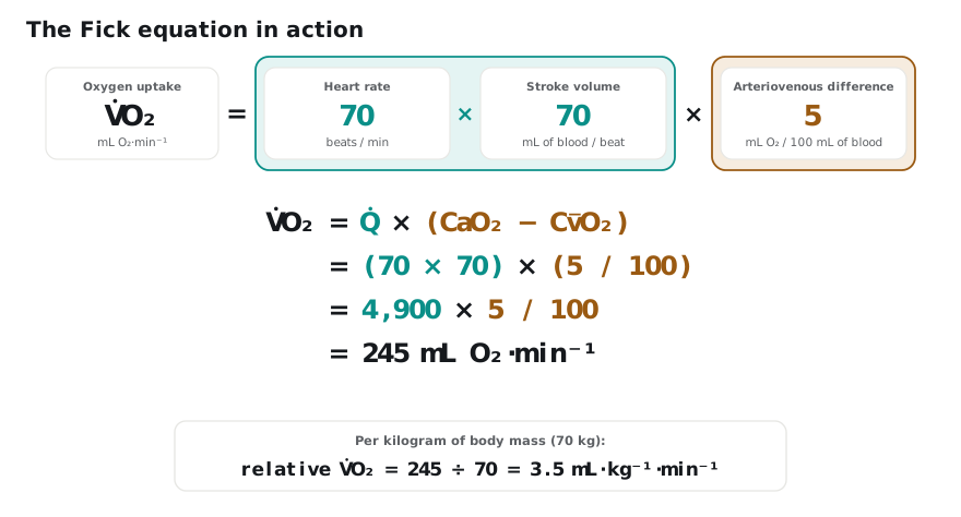
The Fick equation helps us understand what makes up oxygen uptake per unit of time (usually per minute) and what it depends on:
VO₂ = Q × (CaO₂ − CO₂)
This equation tells us that oxygen uptake per minute, VO₂, depends directly on cardiac output, Q, and on the oxygen extracted by our cells from each litre of blood, (CaO₂ − CO₂).
- Q, cardiac output, is simply the volume of blood pumped by the heart per minute. It depends on heart rate (the number of beats per minute) and on the volume of blood ejected with each beat.
- CaO₂ is the arterial oxygen content, i.e. the amount of oxygen in one litre of arterial blood: the blood that the left ventricle sends into the aorta, towards the tissues.
- CO₂ is the oxygen content of mixed venous blood (the bar over the v means “mean”), i.e. the blood returning from all the organs through the veins, which mixes in the right heart and heads back to the lungs through the pulmonary artery.
Their difference, (CaO₂ − CO₂), known as the arteriovenous difference, therefore represents the oxygen taken up by our cells from each litre of blood, at whole-body level, taking all tissues into account (not only the active muscles).
Let us take a concrete example and calculate VO₂ using the Fick equation. Consider a 70 kg person at rest: their heart beats 70 times per minute and ejects 70 mL of blood with each beat (stroke volume), and their tissues take up 5 mL of oxygen from every 100 mL of blood, which is their arteriovenous difference.
Digging deeper
One level down: the ingredients of each term
The Fick equation has only two terms: cardiac output and the arteriovenous difference. Each in turn depends on several ingredients. Let us take them in order.
Cardiac output (Q)
Its two factors (heart rate and the volume of blood ejected with each beat) do not follow the same rules. Maximal heart rate depends mainly on age and cannot be trained: endurance training does not increase it and, if anything, it tends to decrease [16]. What can be trained is stroke volume: the amount of blood the left ventricle sends to the tissues with each beat. It increases with appropriate training [6] and it is what primarily distinguishes athletes from non-athletes [4].
How much blood can the ventricle send out with each beat? This depends first on how much blood it holds just before contracting, i.e. on its filling. Filling depends on the return of blood to the heart, on how quickly the ventricle relaxes between beats and on its compliance, as well as that of the pericardium surrounding it. A larger blood volume could also facilitate this filling [11]. The volume ejected also depends on the force of contraction (contractility, which nervous stimulation increases during exercise [10]) and on the pressure in the arteries, against which the heart must push the blood. Yet what distinguishes athletes is not contractility but a more compliant ventricle, which relaxes faster and fills more: it can therefore send out more with each beat [4].
As effort increases, stroke volume does not follow the same curve in everyone. It was long accepted that it stops increasing at around 40 to 50% of VO₂max [10,11], on the strength of old studies conducted on few subjects and at only two or three intensities: too few data points to draw a solid general conclusion. In reality, the response varies greatly between individuals: in some, stroke volume levels off, or even falls, well before maximal effort; in others, it keeps rising up to VO₂max, as shown by the studies compiled by Vella and Robergs [11]. Fitness level, age, sex, genetics: the explanation is multifactorial.
The arteriovenous difference (CaO₂ − CO₂)
It can be read in two stages: what the arterial blood coming from the left ventricle delivers to the tissues (CaO₂), then what the tissues remove from it, which determines what is left on its return (CO₂).
What the blood delivers: arterial oxygen content, CaO₂. Oxygen in the blood is carried almost entirely by haemoglobin (Hb), a protein contained in red blood cells that binds oxygen:
CaO₂ ≈ 1.34 × [Hb] × SaO₂, plus a small fraction dissolved in plasma
In the expression above, [Hb] is the haemoglobin concentration, in grams per litre, and 1.34 is the number of millilitres of oxygen carried by one gram of haemoglobin [10]. As for SaO₂, this is arterial saturation, i.e. the proportion of that haemoglobin actually loaded with oxygen; it depends on ventilation and on the diffusion of oxygen from the alveoli into the blood [3]. Plasma, for its part, dissolves only about 3 mL of oxygen per litre, whereas 100 mL of whole blood carries about 20 mL, slightly less in women [10]. What matters is therefore not only haemoglobin concentration but the total amount in circulation, which also depends on blood volume, i.e. the total amount of blood circulating in the body: total haemoglobin = [Hb] × blood volume.
What the tissues remove: extraction. For oxygen to be taken up, the blood must first reach the working fibres, the elongated cells that make up muscle. During exercise, blood is redistributed: the arterioles, the small arteries supplying the active muscles, dilate, while those of other organs constrict. At rest, the muscles receive 15 to 20% of cardiac output; during intense exercise, this can rise to 90% [10]. What matters is not only how much blood reaches the muscle, but that this blood goes to the fibres that are consuming oxygen. An experiment reported by Heinonen [16] illustrates this: at maximal exercise, researchers injected the most powerful known vasodilator into the leg artery to force the vessels open. VO₂max did not increase. The fraction of oxygen extracted even decreased: the vessels had also opened towards fibres that were not working. Blood supply no longer matched the needs of the active fibres as precisely [16].
Oxygen must then pass from the blood to the fibre. At the end of the arterioles, blood flows through the capillaries, the finest vessels, which surround each fibre. There, oxygen leaves the red blood cells, crosses the capillary wall and reaches the fibre: this is diffusion, which is easier when the capillary network is dense, the exchange surface is large and the distance to the centre of the fibre is short, which depends on its size [17]. Inside the fibre, two elements take over [17]: myoglobin, a protein similar to haemoglobin that binds oxygen and helps it travel to the mitochondria, and the oxidative capacity of these mitochondria, i.e. the amount of oxygen they are able to use. And the transfer does not stop, because the mitochondria consume oxygen as it arrives: oxygen pressure stays lower in the fibre than in the capillary. It is this gradient that drives it in [5].
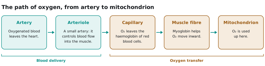
The arteriovenous difference therefore depends on arterial content, on the distribution of blood flow (to the active muscles, then to the working fibres), on diffusion and on the mitochondria: it is not an isolated measure of muscle quality.
Limits and levers
Identifying them to make progress
Limits are the links that cap VO₂max at a given moment. This ceiling is not fixed: which limit weighs most changes with the type of exercise, training status, health and environmental conditions such as altitude, where barometric pressure falls and, with it, the partial pressure of inspired oxygen (hypobaric hypoxia). Oxygen delivery often plays a major role when many muscles are working, without the lungs or the muscles being secondary [3,4,5,16]. A gas exchange measurement (the PEAK option from Twin Peak Lab) sheds light on this overall functioning, but VO₂max alone does not tell where the limit lies in the chain: locating it requires other measurements [2,3].
Levers are what training can act on: loosening a limit when it is trainable (maximal heart rate, for example, is not), or strengthening a point that is already strong. When one limit is loosened, another takes over. And training never works on a single link: stroke volume, the capillary network and mitochondrial content progress together, at different rates; their weight varies with age, starting level and the duration of the effort [6,7,8,16]. Strengthening a single link is not enough anyway: in the mathematical model developed by Wagner [3], we see that degrading a single link makes VO₂max drop markedly, whereas improving it by a comparable amount barely raises it. The idea is therefore to pull these levers in a coordinated way, to get closer to the VO₂max each person can reach given their potential.
Your watch
Now you understand why your watch can get it wrong
Estimating VO₂max, this integrated system involving so many components, seems very difficult from heart rate and external load profiles alone, i.e. power or speed. The watch does not measure gas exchange: it sees neither stroke volume nor what the muscles extract. The error can therefore be large from one person to another [9], which is why it is crucial to measure VO₂max through gas exchange (the PEAK option from Twin Peak Lab) if it is an important parameter for you to know.
In the next article, we will unpack how to stimulate the different components of this integrated system in practice. So you don’t miss it, subscribe to the newsletter.
Arnaud Collet, PhD, CPSS®
References (19)
[1] Wasserman K, Van Kessel AL, Burton GG. Interaction of physiological mechanisms during exercise. J Appl Physiol. 1967;22(1):71–85. doi:10.1152/jappl.1967.22.1.71
[2] Glaab T, Taube C. Practical guide to cardiopulmonary exercise testing in adults. Respir Res. 2022;23:9. doi:10.1186/s12931-021-01895-6
[3] Wagner PD. Determinants of maximal oxygen consumption. J Muscle Res Cell Motil. 2023;44(2):73–88. doi:10.1007/s10974-022-09636-y
[4] Levine BD. VO₂max: what do we know, and what do we still need to know? J Physiol. 2008;586(1):25–34. doi:10.1113/jphysiol.2007.147629
[5] Cardinale DA, Larsen FJ, Jensen-Urstad M, Rullman E, Søndergaard H, Morales-Alamo D, Ekblom B, Calbet JAL, Boushel R. Muscle mass and inspired oxygen influence oxygen extraction at maximal exercise: role of mitochondrial oxygen affinity. Acta Physiol. 2019;225:e13110. doi:10.1111/apha.13110
[6] Rosenblat MA, Granata C, Thomas SG. Effect of interval training on the factors influencing maximal oxygen consumption: a systematic review and meta-analysis. Sports Med. 2022;52(6):1329–1352. doi:10.1007/s40279-021-01624-5
[7] Mølmen KS, Almquist NW, Skattebo Ø. Effects of exercise training on mitochondrial and capillary growth in human skeletal muscle: a systematic review and meta-regression. Sports Med. 2025;55:115–144. doi:10.1007/s40279-024-02120-2
[8] Spina RJ, Ogawa T, Kohrt WM, Martin WH III, Holloszy JO, Ehsani AA. Differences in cardiovascular adaptations to endurance exercise training between older men and women. J Appl Physiol. 1993;75(2):849–855. doi:10.1152/jappl.1993.75.2.849
[9] Molina-García P, Notbohm HL, Schumann M, Argent R, Hetherington-Rauth M, Stang J, Bloch W, Cheng S, Ekelund U, Sardinha LB, Caulfield B, Brønd JC, Grøntved A, Ortega FB. Validity of estimating the maximal oxygen consumption by consumer wearables: a systematic review with meta-analysis and expert statement of the INTERLIVE Network. Sports Med. 2022;52:1577–1597. doi:10.1007/s40279-021-01639-y
[10] Haff GG, Triplett NT, editors. Essentials of Strength Training and Conditioning. 5th ed. National Strength and Conditioning Association. Human Kinetics; 2026.
[11] Vella CA, Robergs RA. A review of the stroke volume response to upright exercise in healthy subjects. Br J Sports Med. 2005;39:190–195. doi:10.1136/bjsm.2004.013037
[12] Hanson NJ, Scheadler CM, Lee TL, Neuenfeldt NC, Michael TJ, Miller MG. Modality determines VO₂max achieved in self-paced exercise tests: validation with the Bruce protocol. Eur J Appl Physiol. 2016;116:1313–1319. doi:10.1007/s00421-016-3384-0
[13] Sietsema KE, Sue DY, Stringer WW, Ward SA. Wasserman & Whipp’s Principles of Exercise Testing and Interpretation: Including Pathophysiology and Clinical Applications. 6th ed. Wolters Kluwer; 2021.
[14] Edvardsen E, Hem E, Anderssen SA. End criteria for reaching maximal oxygen uptake must be strict and adjusted to sex and age: a cross-sectional study. PLoS One. 2014;9(1):e85276. doi:10.1371/journal.pone.0085276
[15] Costa VAB, Midgley AW, Carroll S, Astorino TA, de Paula T, Farinatti P, Cunha FA. Is a verification phase useful for confirming maximal oxygen uptake in apparently healthy adults? A systematic review and meta-analysis. PLoS One. 2021;16(2):e0247057. doi:10.1371/journal.pone.0247057
[16] Heinonen I. Cardiac output limits maximal oxygen consumption, but what limits maximal cardiac output? Exp Physiol. 2025;110(5):666–674. doi:10.1113/EP091594
[17] van der Zwaard S, Brocherie F, Jaspers RT. Under the hood: skeletal muscle determinants of endurance performance. Front Sports Act Living. 2021;3:719434. doi:10.3389/fspor.2021.719434
[18] Michalik K, Danek N, Zatoń M. Comparison of the ramp and step incremental exercise test protocols in assessing the maximal fat oxidation rate in youth cyclists. J Hum Kinet. 2021;80:163–172. doi:10.2478/hukin-2021-0104. These values are taken there from the same authors: J Sports Med Phys Fitness. 2019;59(8):1285–1291.
[19] Day JR, Rossiter HB, Coats EM, Skasick A, Whipp BJ. The maximally attainable VO₂ during exercise in humans: the peak vs. maximum issue. J Appl Physiol. 2003;95(5):1901–1907. doi:10.1152/japplphysiol.00024.2003